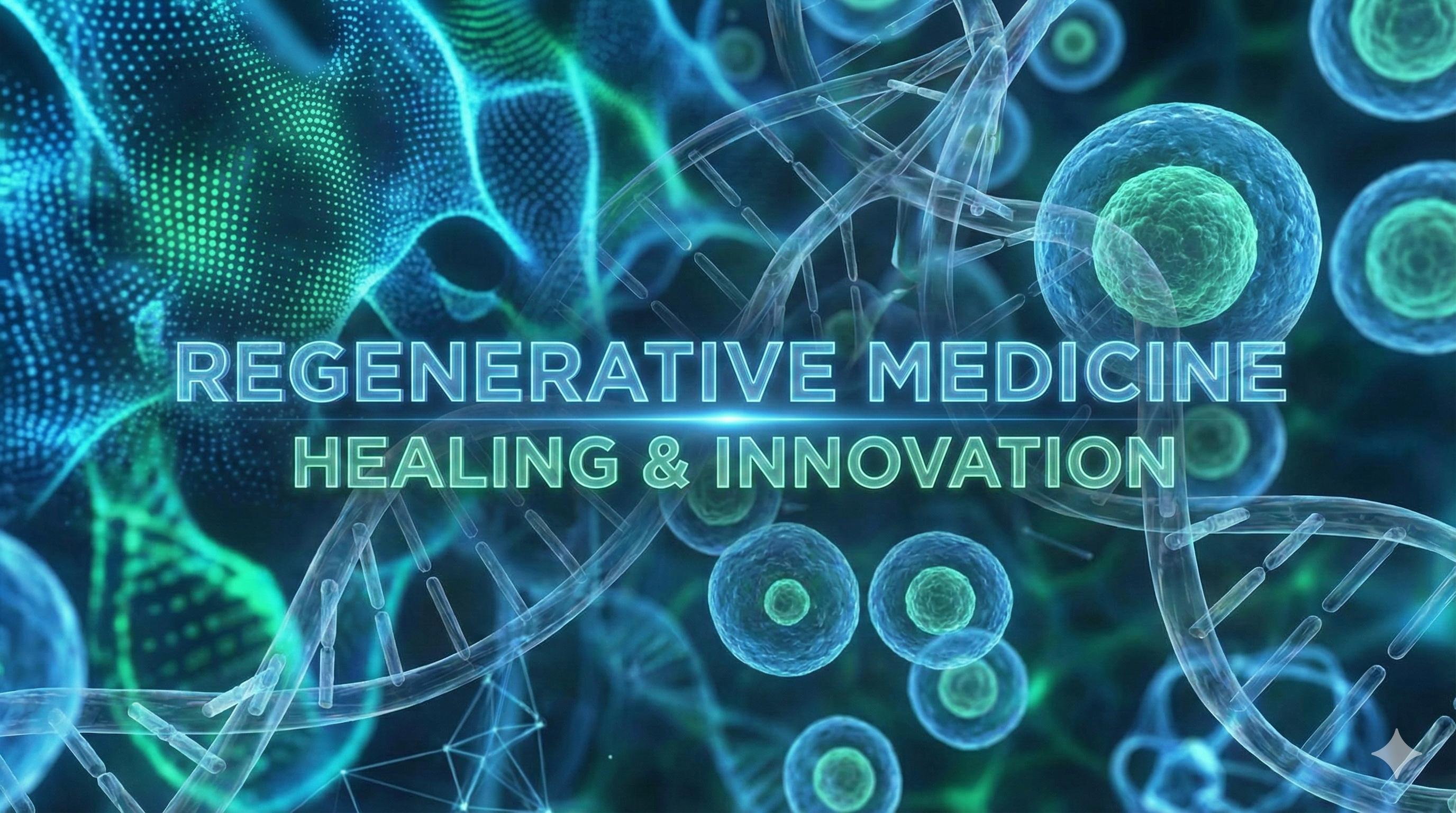
Introduction: The Promise and Challenge of Stem Cells
Regenerative medicine explores the profound possibility of using stem cells to repair or replace tissues damaged by disease, injury, or aging. This concept, once a realm of mythology and legend, is now at the forefront of biological science, offering a potential paradigm shift in treating a wide range of human ailments. However, the path from scientific discovery to clinical application is complex, filled with both immense promise and significant challenges.
The current state of stem cell research can be compared to the early days of personal computing. Looking back at predictions from over 60 years ago, the concept of a personal computer was correct, but the initial design-a massive, complex machine-was entirely wrong. Today, we face a similar situation in regenerative medicine: we have the “right concept” but we are still searching for the correct, safe, and effective stem cell source to replace what may be the “wrong design.”
Scientific progress in the modern era, both in physics and biology, has granted humanity access to technologies of immense power. The development of physics enabled us to harness nuclear energy, a force with the capacity for both great good and terrible destruction. Similarly, advances in biology and genetics have unraveled some of the mysteries of organismal regeneration, leading us into the fascinating world of stem cells. This knowledge, too, presents a dual potential for remarkable healing and for ethically questionable applications—a “dark side” that must be carefully considered. This journey into the building blocks of life is, in many ways, a modern continuation of one of humanity’s oldest dreams: the quest for longevity.
1. The historical and Biological Quest for Longevity
Humanity has always dreamed of achieving longevity. Ancient myths are filled with tales of immortality, from the Ambrosia consumed by the Greek gods to ensure their eternal life, to the Holy Grail sought by the Knights Templar as a source of everlasting youth.
One of the most telling myths is that of Prometheus, who was punished for giving fire to humanity by being chained to a rock where an eagle would eat his liver each day, only for it to regenerate overnight. This ancient story suggests that the Greeks may have astutely recognized the liver’s unique and powerful regenerative capacity, a biological fact we understand well today. Another famous legend is the “Fountain of Youth,” a mythical spring that restores the youth of anyone who drinks its waters. In historical paintings of this legend, it is humorously noted that only women are depicted bathing in the fountain, suggesting a particular interest in the restorative promise of longevity.
This power of regeneration is not just a myth; it is a fundamental part of the biological world.
- Olive trees possess an incredible regenerative potential. Even after being burned or cut down almost completely, they can regrow new branches from the stem and restore the entire tree.
- The salamander is a master of regeneration in the animal kingdom. If it loses a limb, tail, or even the lens of its eye, a cluster of primitive cells called a blastema forms at the site of the injury and regrows the missing part perfectly. This remarkable phenomenon was lost in higher evolution, but the professor suggests that the type of adult stem cells discussed later in this lecture may mimic the function of the blastema on a smaller scale.
2. Regeneration in Humans and Longevity Record
While humans cannot regrow limbs, our bodies are in a state of continuous regeneration. Several key tissues are constantly renewing themselves, a process maintained by reservoirs of adult stem cells.
- Intestine epithelium: The entire lining of the intestine is replaced every 2 days.
- Epidermis: The 1.7-3 square meters of skin covering our bodies is completely renewed every 14 days.
- Erythrocytes (Red Blood Cells): These cells have a lifespan of about 150 days before being replaced.
- Leucocytes (White Blood Cells): These immune cells are turned over much more rapidly, every 4-7 days.
The biological drive for longevity and renewal is also evident in the remarkable lifespans recorded across different species.
- Jeanne Louise Calment (Human): Holds the documented record for human longevity at 122 years. As an anecdote of her vitality, she was reportedly still riding a bicycle at age 100.
- Aldabra Turtle: 256 years
- Japanese Koi (Karp) Fish: 226 years
- Island Mussel – Ming: This ocean quahog holds the record for the longest-living non-colonial animal, reaching an age of 507 years. It was born in 1499 during the Ming dynasty in China, from which it gets its name.
3. The Central Quest: Finding a Pluripotent Stem Cell (PSC)
The central debate in regenerative medicine today revolves around finding the ideal pluripotent stem cell (PSC)—a cell capable of developing into all three primary germ layers of the body (ectoderm, endoderm, and mesoderm). The quest is to identify a source of PSCs that is not only effective but also safe and ethically acceptable for widespread clinical use.
The lecture introduces three main candidates that are at the center of this scientific “war”:
- Embryonic PSCs
- Induced PSCs
- Adult tissue-derived PSCs
The following sections will evaluate each of these candidates, exploring their biological origins, potential therapeutic uses, and the significant pitfalls associated with them.
Part 1: Embryonic and Induced Pluripotent Stem Cells
Understanding embryo-derived and induced pluripotent stem cells is strategically crucial, as they were the first types of PSCs discovered and have historically dominated the research landscape. However, they are fraught with significant scientific and ethical challenges that have, to date, prevented their widespread clinical application. Grasping these limitations is essential to appreciate the ongoing search for safer and more viable alternatives.
1. Embryo-derived Pluripotent Stem Cells (ESCs)
Embryonic Stem Cells (ESCs) are derived from the earliest stages of development. Following fertilization, a single-celled zygote develops over several days into a blastocyst. This structure contains an outer layer that will form the placenta and an inner cell mass (ICM), a small cluster of cells that are naturally pluripotent and destined to form the entire organism.
This is the source of the core ethical controversy. The Catholic view holds that life begins at fertilization, making the destruction of a blastocyst to harvest its ICM morally unacceptable. In contrast, in many other major religions like Judaism or even Islam, they actually consider that life begins at the time of implantation in the uterus. This distinction is critical, as ESCs are derived from the pre-implantation blastocyst, often using surplus embryos from in-vitro fertilization (IVF) clinics that would otherwise be discarded.
Despite the initial excitement, research on ESCs has revealed significant problems that have severely limited their therapeutic potential.
- Risk of teratoma formation: When ESCs are transplanted, there is a high risk that they will form a teratoma, a monstrous tumor containing a disorganized mix of tissues like hair, teeth, bone, and muscle. This is a primary safety concern.
- Ethical and religious controversy: The use of human embryos remains a deeply divisive issue, creating legal and social barriers to research and application.
- ★ Lack of significant scientific progress: This is arguably the most critical practical issue. Despite being studied for over 20 years, ESCs have not led to significant clinical breakthroughs. Early clinical trials were initiated but were subsequently stopped due to safety and efficacy concerns. This decades-long stagnation, despite enormous initial hype and funding, has led many in the field to question whether ESCs will ever become a practical, widespread therapeutic solution, shifting focus toward alternative strategies.
- Problem with histocompatibility: ESCs are not a genetic match to a patient. They possess HLA antigens from both biological parents and would be rejected by a recipient’s immune system, just like an organ from an unmatched donor.
- Availability of alternative strategies: As the field has advanced, other methods to obtain PSCs have been developed, reducing the sole reliance on controversial embryonic sources.
2. Nuclear Transfer-derived Pluripotent Stem Cells (Therapeutic Cloning)
To overcome the histocompatibility problem of ESCs, Dr. John Gurdon developed the nuclear transfer process, a technique also known as therapeutic cloning. This groundbreaking work earned him a Nobel Prize.
The process involves several key steps:
- An oocyte (egg cell) is obtained from a donor, and its nucleus is surgically removed (enucleated).
- The nucleus from one of the patient’s own somatic cells (e.g., a skin cell) is isolated and transferred into the enucleated oocyte.
- This reconstructed cell, termed a “clonote,” contains the patient’s nuclear DNA within the rich developmental cytoplasm of the oocyte.
- The clonote is stimulated to develop into a blastocyst, from which PSCs can be isolated from the inner cell mass.
The primary advantage of this method is that the resulting stem cells are a near-perfect histocompatibility match to the patient, as they contain the patient’s nuclear DNA. This allows for the creation of “custom-made” cells that should not be rejected by the immune system.
However, this technology has a “dark side”: its potential for abuse in reproductive cloning. If the clonote is implanted into a surrogate mother’s uterus instead of being used to derive stem cells, it could theoretically develop into a full organism—a clone of the nucleus donor. The most famous example of this is Dolly the sheep, the first mammal cloned from an adult cell. It is important to note that Dolly lived only half as long as her genetic mother and developed premature disorders, highlighting the dangers and imperfections of the process.
This potential for human cloning has raised speculative fears of creating armies of “Star Wars warriors” and has also fueled scientific projects aimed at de-extinction, such as cloning a mammoth using a nucleus from a frozen carcass and an elephant oocyte. The scientific community has widely condemned the practice of human reproductive cloning. The publication of two papers in the journal Cell reporting the first successful steps of human therapeutic cloning was met with ethical condemnation, underscoring the serious reservations held by researchers.
Finally, even these “custom-made” cells are not entirely without immune issues. While the nuclear DNA matches the patient, the mitochondrial DNA comes from the oocyte donor. It has been shown that mismatched antigens encoded by this mitochondrial DNA can still trigger an immune rejection of the transplanted cells. This is because mitochondria contain their own small genome which encodes proteins. If these proteins differ between the oocyte donor and the patient (nucleus donor), the patient’s immune system can recognize them as foreign antigens, leading to rejection of the very cells designed to be a perfect match.
3. Induced Pluripotent Stem Cells (IPSCs)
A major breakthrough came with the development of induced Pluripotent Stem Cells (iPSCs) by Dr. Shinya Yamanaka, for which he shared the Nobel Prize with Dr. Gurdon. This revolutionary method creates pluripotent cells without the need for embryos or donor oocytes, thereby circumventing the major ethical controversies.
The process involves:
- Taking a patient’s own somatic cell, such as a skin fibroblast.
- Exposing the cell to a cocktail of specific genes, known as the “Yamanaka factors” (e.g., Oct-4, Klf-4, c-myc). This can be done by introducing the genes themselves or their corresponding mRNA or proteins.
- This exposure forces the adult cell to de-differentiate, essentially rewinding its developmental clock back to a primitive, pluripotent state.
Despite its ethical advantages, iPSC technology is plagued by its own set of serious problems, particularly concerning safety.
- Immune response to autologous iPSCs: Even when derived from the patient’s own cells, iPSCs can sometimes trigger an immune response upon transplantation.
- Genomic instability: During the extensive cell culture (passaging) required to grow iPSCs, they tend to acquire chromosomal abnormalities and other genetic mutations.
- Risk of insertional mutagenesis: The vectors (often viruses) used to deliver the Yamanaka factors can insert themselves into the cell’s DNA randomly. This can damage essential genes, such as disabling anti-oncogenes that protect against cancer.
- Variability among iPSC clones: Different iPSC clones derived from the same starting cells can behave very differently, making standardized therapeutic production difficult.
- ★ Safety concerns: This is the most critical barrier to clinical use. Like ESCs, iPSCs have a very high risk of forming teratomas after transplantation. The professor makes a powerful point: no researcher who works with iPSCs, including Yamanaka himself, would consent to having these cells injected into their own body due to this known and significant cancer risk.
4. Comparison of Pluripotent Stem Cell Sources
The following table summarizes the key features and drawbacks of the three major PSC sources discussed so far.
| Feature | ESCs (from Fertilization) | PSCs (from Cloning) | iPSCs (from Reprogramming) |
|---|---|---|---|
| Risk of Teratomas | High (+++) | High (+++) | High (+++) |
| Histocompatibility Problem | Yes (+) | Potential (+/-) | Yes (+) (for allogeneic use) |
| Requires Donor Ovum | Yes (+) | Yes (+) | No (-) |
| Ethical Reservations | Yes | Yes/No* | No |
This problem is differently considered by various major religions of the world. A number of religions potentially accept therapeutic cloning (for example Judaism, Islam, Buddhism), but the vast majority have reservations about reproductive cloning.
Given that the leading PSC candidates—ESCs, cloned PSCs, and iPSCs—are all critically flawed by the shared risk of teratoma formation and burdened with significant ethical and logistical hurdles, the scientific community began an urgent search for a safer alternative: a pluripotent stem cell that might naturally reside within our own adult tissues.
Part II: Pluripotent Stem Cells from Postnatal Tissues
Given that the leading pluripotent stem cell (PSC) candidates are plagued by safety and ethical concerns, the focus of regenerative medicine has increasingly shifted toward a groundbreaking idea: that true, safe PSCs might naturally reside in our bodies long after birth. The strategic importance of this search cannot be overstated. If such a population of cells exists, they would bypass the profound ethical dilemmas of ESCs and iPSCs and would likely possess innate biological mechanisms to prevent tumor formation, addressing the single greatest safety concern in the field. The central question became: “if such pluripotent stem cells exist in postnatal tissues…which could give rise to all three germ layers?”
1. The Debate and The Old “Stem Cell Plasticity” Concept
The acknowledgment of pluripotent stem cells in adult tissues has been a two-decade struggle for several reasons:
- They are extremely rare, making them very difficult to isolate and study.
- Much of the early evidence was indirect and not performed at the single-cell level, making it easy to dismiss.
- There was an overestimated hope that ESCs and iPSCs would quickly move to the clinic, diverting attention and funding.
- There has been significant competition from powerful ESC and iPSC research lobbies.
- The field was damaged by the promotion of a false concept known as “stem cell plasticity.”
The concept of trans-dedifferentiation (or plasticity) was the belief that an adult, tissue-committed stem cell—like a hematopoietic stem cell (HSC) from the bone marrow—could spontaneously change its fate and become a cell of a completely different organ, such as a cardiomyocyte (heart muscle) or a neuron. However, the professor clearly states that this concept was not confirmed. The sensational papers supporting it, though never formally retracted, were found to be based on flawed interpretations and could not be replicated.
2. Alternative Explanations for Regenerative Effects
Given that plasticity was not the answer, the professor’s team postulated three alternative explanations for the positive therapeutic effects that were sometimes observed when bone marrow cells were used to treat non-blood-related disorders.
Explanation 1: Paracrine Effects (Soluble Factors)
The first hypothesis was that stem cells act as a “factory” for beneficial molecules. Instead of turning into new tissue themselves, they release a cocktail of growth factors, cytokines, chemokines, and bioactive lipids. These soluble factors then act on the surrounding damaged tissue to promote healing. The professor references his own paper, published over 20 years ago, demonstrating that human CD34+ cells (a population enriched for hematopoietic stem cells) secrete numerous factors that protect damaged tissue by inhibiting apoptosis (programmed cell death) and promoting vascularization (the formation of new blood vessels).
Explanation 2: Extracellular Microvesicles and Exosomes
The second explanation involves tiny vesicles shed by stem cells. These include microvesicles, which bud off from the cell surface, and exosomes, which originate from internal compartments. These vesicles are loaded with biological cargo, including mRNA, microRNA, and proteins. They can fuse with other cells and transfer this cargo, effectively reprogramming the target cells to prevent cell death and improve their function. The professor notes that his 2006 paper was the first to demonstrate this mechanism of horizontal transfer of biological information via vesicles.
Explanation 3: Presence of Dormant Pluripotent Stem Cells
This was the professor’s primary hypothesis: that the regenerative effects were not due to plasticity or solely paracrine effects, but to the existence of a very rare, primitive population of pluripotent stem cells already residing in the bone marrow. He proposed two possible developmental scenarios:
- Scenario 1: PSCs exist only in the embryo, give rise to tissue-committed stem cells (TCSCs), and then disappear entirely.
- Scenario 2: After creating the TCSCs, a backup population of PSCs survives into adulthood. To prevent them from forming teratomas, they are kept in a deeply quiescent, “locked” state.
3. The Discovery of Very Small Embryonic-Like (VSEL) Stem Cells
The candidate for this dormant adult PSC population was discovered and named Very Small Embryonic-Like (VSEL) Stem Cells. These cells were identified in adult bone marrow, human cord blood, and other tissues.
Based on electron micrographs, they have distinct physical characteristics:
- They are very small, with a diameter of approximately 6 micrometers, making them slightly smaller than a 7-micrometer red blood cell.
- They have a very large nucleus that occupies most of the cell volume. Their large nucleus is filled with open euchromatin, a loosely packed form of DNA that indicates high transcriptional potential, which is a hallmark of primitive, undifferentiated cells. This contrasts sharply with the mature Hematopoietic Stem Cell (HSC), whose nucleus contains patches of condensed heterochromatin, a tightly packed, transcriptionally inactive form of DNA.
- There is only a very tiny rim of cytoplasm surrounding the nucleus.
- They feature structures called nuancesomes around their mitochondria, a feature characteristic of the germline.
The professor notes that VSELs were missed by researchers for decades because their small size caused them to be discarded along with red blood cells and cellular debris during standard cell sorting procedures.
4. Evidence for VSEL Pluripotency
Multiple lines of molecular evidence support the claim that VSELs are indeed pluripotent.
- Molecular Signature: Analysis of their gene expression (shown as a heat-map) reveals a fingerprint very similar to that of embryonic stem cells. They actively express a suite of genes associated with pluripotency (like Oct-4 and Nanog), epiblast development, and germline specification.
- Oct-4 Expression: The Oct-4 promoter—the switch that turns the master pluripotency gene on or off—is hypomethylated (un-methylated) in VSELs, meaning it is active, just as it is in ESCs. In contrast, the Oct-4 promoter in HSCs is heavily methylated and thus silenced.
- Open Chromatin: Chromatin immunoprecipitation (ChIP) assays confirm that the Oct-4 promoter in VSELs has an open chromatin structure, marked by acetylated histones (H3Ac). This indicates that the gene is primed and ready for transcription.
Furthermore, analysis shows that VSELs contain a normal, diploid amount of DNA and are viable, proving they are a distinct cell population and not merely apoptotic debris.
5. VSELs in the Body: Location and Mobilization
VSELs are not confined to the bone marrow; they have been isolated from numerous other organs, including the lungs, brain, kidney, pancreas, and liver.
★ One of the most important findings is that the number of VSELs present in tissues decreases significantly with age. This decline may be linked to the reduced regenerative capacity and increased frailty associated with aging.
VSELs are not static. During times of stress or injury, they are mobilized from their tissue niches (especially the bone marrow) and released into the bloodstream. They circulate throughout the body and are guided to sites of injury by a chemical distress signal, specifically an SDF-1 gradient, which acts on the CXCR4 receptor present on the surface of VSELs.
Mobilization of VSELs into the peripheral blood has been documented in humans in response to several clinical situations:
- Acute Myocardial Infarction (Heart Attack)
- Stroke
- Skin Burn Injuries
- Crohn’s Disease
- Psychiatric DIsorders (specifically during acute psychotic episodes)
This evidence supports the professor’s hypothesis that VSELs are a natural, mobile reserve population activated by injury to assist in regeneration. This system functions as a sort of internal repair crew, analogous to the blastema in a salamander, but operating on a much smaller scale. Over the years, other laboratories have identified similar primitive cell populations and given them different names (MASC, Muse, MAPC, MIAMI, etc.). The professor believes that these are likely overlapping discoveries of the same biological system, with VSELs representing the apex cell at the top of this adult stem cell hierarchy.
Part III: The Mechanism and Implications of VSELs
This final section delves into the sophisticated biological mechanisms that regulate VSELs, keeping them dormant and safe. It further explores their profound implications for some of biology’s biggest questions, including the processes of aging, the origins of cancer, and the future direction of regenerative medicine.
1. The Key to Quiescence: Erasure of Genomic Imprinting
Understanding why VSELs are naturally quiescent (dormant) was the crucial puzzle that needed to be solved before they could be expanded in the lab (ex vivo) for therapeutic use. A key safety feature of VSELs is that they do not meet the official in vivo criteria for pluripotency: they do not cause teratoma formation when injected, nor do they contribute to blastocyst complementation. This innate safety is what makes them so attractive compared to ESCs and iPSCs.
The professor’s central hypothesis for their quiescence connects them to the germline—the lineage of cells that give rise to gametes (sperm and eggs). The mechanism lies in a process called somatic imprinting.
- Somatic imprinting is an epigenetic mechanism that silences one copy (either maternal or paternal) of certain developmentally important genes.
- The primary example is the Igf2-H19 locus. In normal somatic cells, the paternal copy of the Igf2 gene (which promotes growth) is expressed, while the maternal copy is silenced. This maintains a balanced level of growth signaling.
- However, during the development of the germline, in cells known as primordial germ cells (PGCs), this somatic imprint is completely erased. This is a crucial step in “resetting” the genetic slate for the next generation.
★ The professor’s key discovery was that VSELs, like primordial germ cells, have an erased somatic imprint at the Igf2-H19 locus. The biological consequence of this is profound: because the imprinting that allows the paternal Igf2 gene to be expressed is gone, Igf2 is silenced in VSELs. This makes the cells deaf to powerful growth signals from IGF-1 and Insulin. This unresponsiveness to major growth factor pathways is the molecular lock that keeps VSELs dormant and safely prevents them from proliferating uncontrollably and forming teratomas.
2. VSELs: Implications for Aging and Longevity
Building on this discovery, the professor proposes a theory for aging: VSELs are “burned during life in the metabolic fire” of the Insulin and Insulin-like Growth Factor (IIS) signaling pathway.
There is a well-established connection between the IIS pathway and lifespan across the biological kingdom:
- Down-regulation of IIS signaling, achieved through caloric restriction or genetic mutations, is known to extend lifespan and lower cancer risk in organisms from yeast to mice.
- Conversely, high IIS signaling, often resulting from a high-calorie diet, is associated with a decreased lifespan and an increased cancer risk.
The “Laron Dwarf” mice provide a compelling example:
- These mice have a genetic mutation in the Growth Hormone (GH) receptor, which results in very low levels of IGF-1.
- As a result, they are dwarfs, but they live 25-65% longer than normal mice and are highly resistant to developing cancer.
- Crucially, the professor’s lab discovered that these long-living mice have a preserved population of VSELs in their bone marrow, even into old age, whereas normal old mice have very few.
This suggests that low IIS signaling preserves the body’s reservoir of VSELs, contributing to a longer, healthier life. This finding also extends to fertility. Evidence suggests that VSELs in the ovaries can serve as precursors for new oocytes. Supporting this, Laron dwarf mice remain fertile at 2 years of age, an age when normal mice are long past their reproductive window.
3. VSELs: Implications for Cancerogenesis
The discovery of VSELs provides a modern molecular basis for the historic “embryonal rest theory of cancer,” first proposed by Julius Cohnheim in 1875. This theory suggested that cancers arise from dormant embryonic remnants left behind in adult tissues.
Several data points support a modern version of this theory, implicating the germline/VSELs as a potential origin of cancer:
- The existence of classical germline tumors like seminomas and teratomas.
- The expression of Cancer-Testis (C/T) Antigens in many types of cancer. These antigens are normally expressed only in the germline, but they are aberrantly activated in tumors. VSELs also express C/T antigens.
- The expression of the master pluripotency marker Oct-4 in several tumor types.
The link between imprinting, growth signaling, and cancer is powerfully illustrated by Beckwith-Wiedemann Syndrome. In this condition, a genetic error causes a Loss of Imprinting (LOI) at the Igf2-H19 locus. This leads to the expression of both copies of the Igf2 gene, causing massive overexpression of the growth factor. The result is fetal overgrowth and a very high risk of developing pediatric sarcomas. This shows what happens when the epigenetic “lock” that keeps VSELs quiescent is broken.
This leads to the professor’s final conclusion about the “Yin and Yang” relationship between VSELs and the IIS pathway:
- High IIS signaling (Yang) depletes the VSEL pool, leading to accelerated senescence (aging) and an increased risk of cancer.
- Low IIS signaling (Yin) preserves the VSEL pool, leading to prolonged longevity and a lower risk of cancer.
4. Final Conclusions
The lecture concludes with a summary of the key takeaways from this exploration of regenerative medicine.
- The quest for a pluripotent stem cell that is safe, effective, and ethically accepted for clinical use still continues.
- Most of the current therapeutic benefits seen in adult stem cell therapies for organ damage are likely due to paracrine effects (secreted factors) and cellular reprogramming by microvesicles and exosomes, rather than direct tissue replacement.
- Evidence is accumulating that developmentally early stem cells, such as VSELs, reside in adult tissues. As new strategies to expand them are developed, they may become a viable option for clinical use.
- ★ An important aspect of anti-aging strategies will be to increase the robustness of the body’s resident stem cell populations through pharmacology, diet, or physical activity.
Leave a Reply